Monthly Archives: January 2015

A Talk With a Cancer Patient

2015/01/img_0514.jpg
This past week, I had the privilege to talk with and learn from a survivor of cancer. My interviewee was a seventy-seven year old man who had prostate cancer. While he is in remission, I could tell this was still a very sensitive and painful topic to discuss. Two years prior, he had joined a new health care system, and had gone to a routine medical checkup for the first time in a few years. On a routine blood test, a PSA level was checked, and it was determined that it was elevated. The PSA, or prostate specific antigen was eleven. He told me this meant the chance of him having prostrate cancer was over fifty percent. He then underwent a CT Scan, a bone scan, and eventually a prostate biopsy. The prostate biopsy “was uncomfortable,” he remarked. He joked, “he had no symptoms before the biopsy, only after.” After it was discovered that he had cancer, he alternated between denial and “I am going to beat this” attitude. Admittedly, he told me, that he was scared of the dreaded c-word. After he was diagnosed, he told me that he was given a choice; surgery, or radiation and hormone therapy. He was reluctant to have surgery in fear of lifelong incontinence, so he chose hormone therapy with radiation. My survivor received external beam radiation, five days a week for a nine week period and he started a hormone therapy called Lupron therapy. He told me that the radiation therapy targeted the cancer cells in his prostate, and the Lupron stopped the release of testosterone. This slows or stops the growth of cells including his cancer cells which are testosterone dependent. Initially, his travel plans with his wife had to be altered to allow for the nine weeks of radiation therapy. He associated the treatment with his extreme fatigue, which prohibited him from enjoying his everyday lifestyle. My interviewee said that he was grateful for his wife in helping him, and for all the support from his family and friends, while struggling with not wanting to be a burden to them. He grew closer to a friend who had previously survived prostate cancer, but did not feel comfortable talking about it to most of his other friends. He focused more on the present tense than the future, enjoying each moment. Most of his difficultly centered upon the “what ifs” and struggling to deal with the possibility of the worst possible outcome. In regard to addressing common misconceptions about cancer, he replied, “people believe that you have no voice in your options for treatment. However, the most important thing is to become educated in your treatment options, and discuss them openly with your physician. The goal is to work with your doctor, “not follow blindly.” He shared personal advice as well: “appreciate every day; allow yourself to be inspired and inspire others. No matter what circumstance you are in, keep a positive outlook on the future and don’t dwell on the ‘what-ifs.'”

After talking with a cancer patient, I developed a strong admiration for all of those who endure the hardships of cancer. His views on life drastically changed mine, reminding me to focus on the present and not dwell on the past, or the future. Reading the text book and classroom discussions gave me a third party detailed view on cancer, but actually speaking to somebody who had experienced cancer firsthand deepened both my knowledge and perception of cancer, as well as being an august experience. I am grateful to my interviewee for all that he shared with me, and I will remember it forever.

About Prostate Cancer

According to E.O. Wilson’s Life on Earth cancer is “a disease in which abnormal cells divide uncontrollably and invade other tissues. A cancer can occur anywhere in the human body, and then spread to other tissues via metastasis.

My cancer patient developed prostate cancer, which affects the prostate gland in males. The prostate gland is a part of the male reproductive system located between the bladder and the rectum. Prostate cancer, if it becomes metastatic, can spread throughout the body, even affecting vital organs such as the liver, lung, brain and heart. Luckily, my interviewee’s cancer did not become metastatic, and was contained to the prostate.

The cause of changes that lead to prostate cancer is not known by medical professionals, however, medical researchers now understand the changes in DNA that can create cancerous prostate cells.

Contrary to popular belief, genetics are only responsible for approximately 5-10% of all prostate cancers. In these cases, genetic inheritance of three subsets of gene mutations lead to a higher rate of prostate cancer.

One of these gene mutations is RNASEL, derived from HPC1. HPC1 kills tumors when they appear in the prostate glands. The mutation of RNASEL allows the irregular cells to last longer, increasing the chance of developing prostate cancer.

Another two genes that can harmfully mutate include BRCA1 and BRCA2. These genes suppress tumors by fixing mistakes in DNA synthesis. Although mutations in these genes are uncommon, they account for a number of prostate cancers, as well as breast and ovarian cancers.

MSH2 and MLH1 are another two genes that correct irregularities in DNA. Men who inherit mutations in this gene develop the condition Lynch syndrome, also called hereditary nonpolyposis colorectal cancer (HNPCC). Men with Lynch syndrome are at a higher risk at developing prostate, colorectal, small intestine, stomach, gallbladder, and brain cancers.

In addition to inherited genes that may cause cancer, some scientists hypothesize that DNA mutations or high production of substances can attribute to increased risk of developing cancer. Increased androgen level, such as large amounts of testosterone, have been proven to promote cell growth, perhaps contributing to the cell growth of cancerous cells in the prostate.

Another hormone, insulin-like growth factor-1 (IGF-1), perhaps can correlate to an increased risk of developing prostate cancer. IGF-1 is another hormone that regulates cell growth that some studies have related to cancer growth, however, additional studies are yet to connect IGF-1 and prostate cancer.

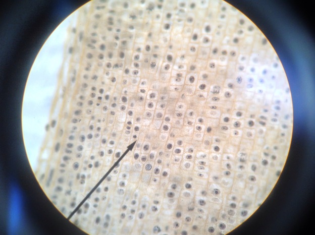
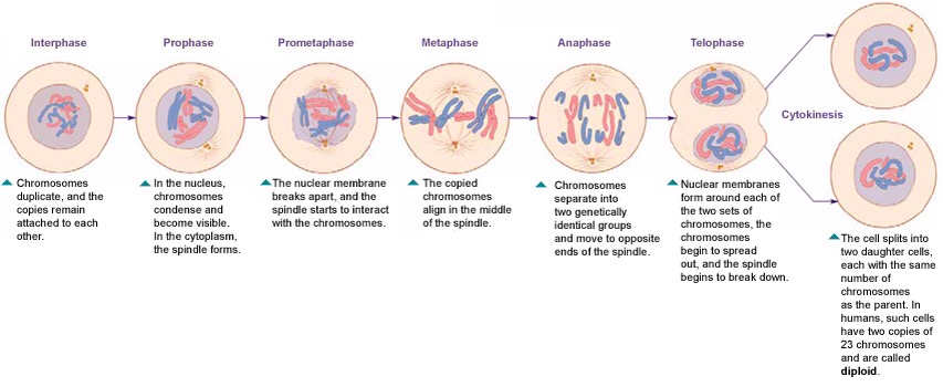
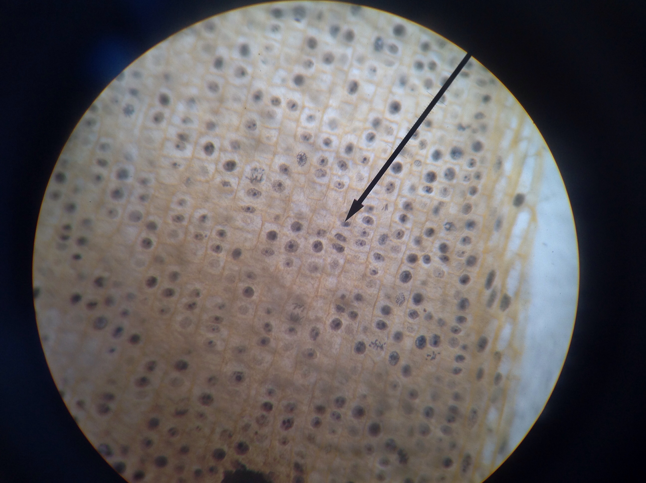
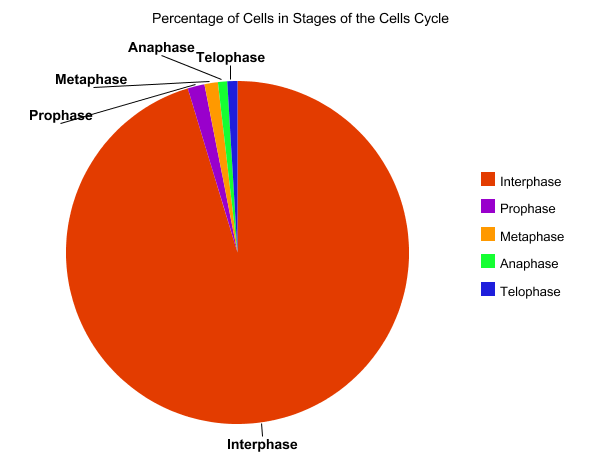
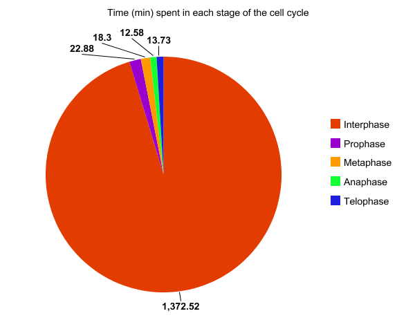
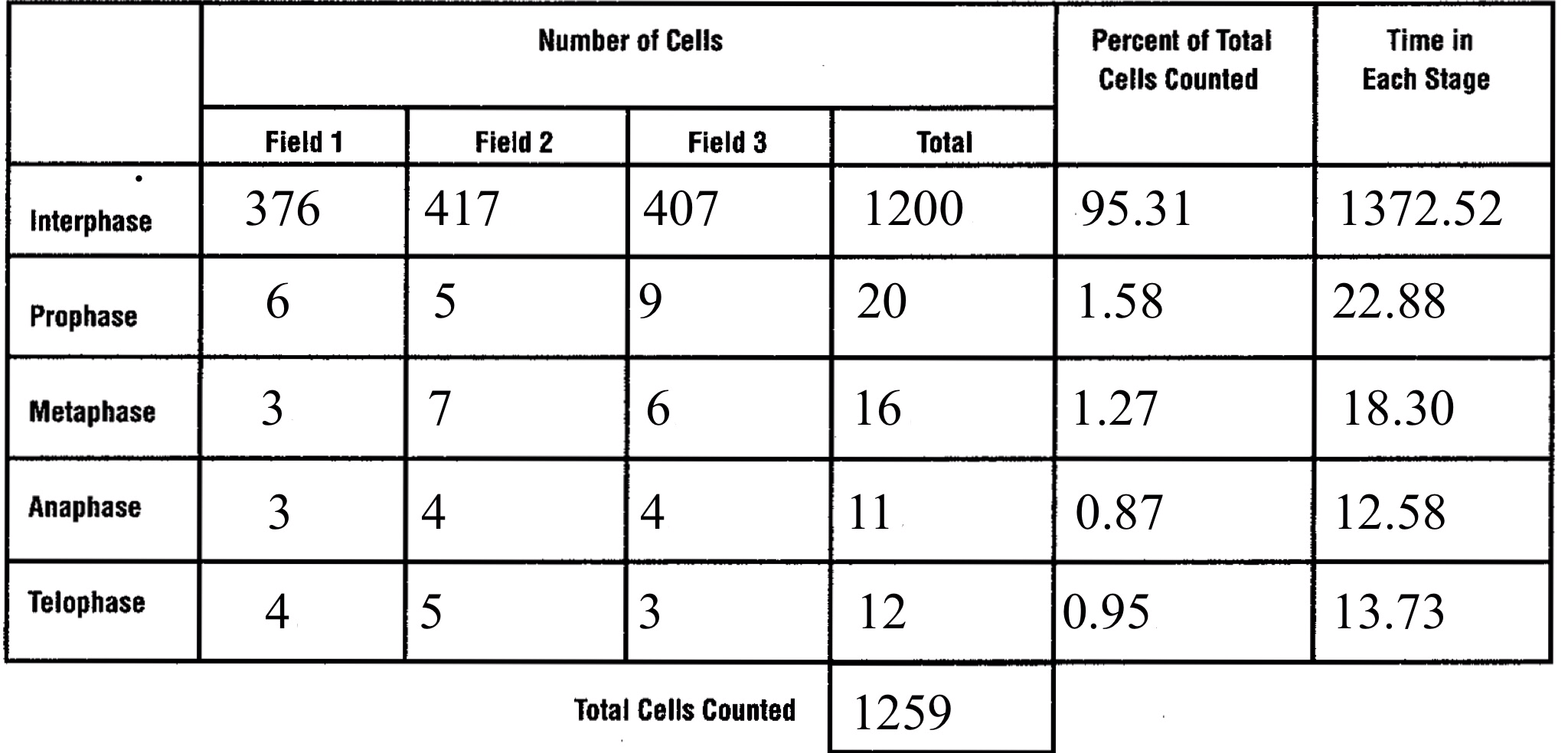
In prostate cancers, the cell cycle plays an important role. When DNA replicates during the S stage of interphase, mutations in genes described above can pass through the cell cycle, producing new cancerous cells, which can then uncontrollably divide as tumors. Other proteins that interact with cells during different stages of the cell cycle affect, modulating ARs. These include “G0 (RB), G1 to S phase (cyclin D1, cyclin E, Cdk6), or G2 (Cdk1)” (Balk-Knudsen). Errors in the modulation of ARs could possibly cause a cancer to develop.

To treat prostate cancer, medical professionals use many techniques. The patient usually can consider which path to take, dependent upon the patient’s age and life span, other health problems, the stage of the cancer, and medical opinions.
Patients with stage one prostate cancer are recommended to undergo “active surveillance”, and treatments include radiation therapy involving either external beams or brachytherapy, and radical prostatectomy.

Patients with stage two prostate cancer have the same treatment options as stage one prostate cancer patients, however, a majority of radiation therapies also include hormone therapy as well.

Patients with stage three prostate cancer usually also have the same options as stage two patients, but it is recommended by medical professionals to have radiation, then a radical prostatectomy.

Finally, patients with stage four prostate cancer unfortunately usually cannot partake in the standard cancer treatments. Treatment for stage four patients include hormone therapy, surgery to remove pain in the bladder or rectum. Bone metastatic development treatment, such as denosumab, a drug also used to cure osteoporosis, or “radiopharmaceutical such as strontium-89, samarium-153 or radium-223.” (Cancer.org)

As we do not know a definite cause for prostate cancer, there are not strong evidence of prevention, however many other ways to prevent cancers remain true for prostate cancer.
The American Cancer Society suggests that one eats 2 1/2 cups of assorted fruits and vegetables daily, one maintains a healthy weight and diet as well. Additional studies have shown that some medicines can also decrease the rate of developing prostate cancer. Early studies show that supplements of Vitamin E and selenium lower the rate of prostate cancer in males. Furthermore, “5-alpha reductase inhibitors [treat]!prostatic hyperplasia (BPH), a non-cancerous growth of the prostate” (cancer.org) These medicines include Finasteride, marketed as Proscar, and Dutasteride marketed as Avodart may decrease the risk of developing prostate cancer. Other studies suggest that prolonged aspirin intake could also decrease the rate of prostate cancer, but further studies are required.

In 2014, it is estimated that another 233,000 men in the U.S. will develop prostate cancer, and approximately 1,112,000 people will be diagnosed in the world. Of these numbers, 29,480 men in the U.S. Will die of prostate cancer.

Physicians whom help treat prostate cancer include:
–Urologists, or a surgeon that specializes in surgery of the urinary tract and the male reproductive system.
–Radiation oncologists, a physician who treat cancer with radiation therapy, and
–Medical oncologists, a doctor who treats cancer with chemotherapy or hormone therapy.

Sources
National Cancer Institute
http://www.cancer.gov/cancertopics/types/prostate
National Cancer Association
http://www.cancer.org/cancer/prostatecancer/detailedguide/prostate-cancer-treating-general-info
http://www.cancer.org/cancer/prostatecancer/detailedguide/prostate-cancer-treating-by-stage
http://www.cancer.org/cancer/prostatecancer/detailedguide/prostate-cancer-prevention
http://www.cancer.org/cancer/prostatecancer/detailedguide/prostate-cancer-what-causes
World Cancer Research Foundation
http://www.wcrf.org/int/cancer-facts-figures/worldwide-data
Genetics Home Reference
http://ghr.nlm.nih.gov/condition/lynch-syndrome
Balk, Steven and Knudsen, Karen : National Center For Biotechnology Information
http://www.ncbi.nlm.nih.gov/pmc/articles/PMC2254330/#__sec6title

Mitosis Lab

Abstract

In this experiment, I investigated slides of root tips and analyzed the phases of mitosis and the relative duration of each phase in the meristem root tissue. An organism’s growth is delicately controlled by regulating the cell cycle. Mitosis is the cell cycle process in which nuclear division and cytokinesis form two identical daughter cells.

Introduction and Background

In the early 1880s, the German anatomist Walther Flemming observed the fibrous structures of spindle fibers and created the term mitosis, from the Greek word for thread, to describe the actions of spindle fibers in a dividing cell.
To study the process of mitosis, I examined cells from the meristem root tissue of an onion, in which many cells are undergoing the process of mitosis. Mitosis is the process of nuclear division and cytokinesis to form two identical daughter cells.
Mitosis consists of the following stages: interphase (although not technically considered part of mitosis, it is the precursor to mitosis), in which the chromosomes duplicate and the cell readies itself for mitosis; prophase, in which chromosomes condense and spindle fibers form; prometaphase, when the nuclear membrane disintegrates; metaphase, when the chromosomes line up one the middle of the spindle fibers with the centromere attached to the spindle; anaphase, in which the chromosomes separate and migrate towards the asters; and telophase, when nuclear membranes begin to form around the chromosomes on both sides of the cell. During telophase, the chromosomes condense and the cell prepares for cytokinesis, in which the cell splits into two identical daughter cells. Interphase and the stages of mitosis are seen in Figure 1 below.

Figure 1:

2015/01/img_0489.jpg

I used onions in this experiment due to the high percentage of cells that undergo mitosis in an onion’s apical meristem root tissue. This tissue is located towards the end of the root, near the root stem. The location of the apical meristem root tissue is displayed in Figure 2 below.

I used whitefish blastula in this experiment because of its rapid rate of nuclear division from fertilization. This rapid division allows one to visualize spindle fibers very clearly in the cell.

Figure 2:

2015/01/img_0490.png

The duration of the stages of mitosis can be determined by counting the number of cells in mitosis at a given time and multiplying that value by the average duration of mitosis in an onion, which is approximately twenty-four hours.

I hypothesized that a majority of cells would be in interphase, because of the time required to synthesize new DNA molecules during the S stage.
Materials and Procedure

Materials List

–1 Light Microscope, outfitted with a 10X eyepiece and 4X, 10X, and 40X objective lens
–Prepared Microscope Slide of 3 Onion Root Tips
–Prepared Microscope Slide of Whitefish Blastula

Procedure

Part One: Observing Mitosis in Plant and Animal Cells

1. I brought the microscope into focus on the lowest 4X setting with the onion root tip in view, making sure the diaphragm was at its widest setting.
2. I located the meristem root tissue of the onion root tip and switched the microscope to the 40X measure, in which cells were visible.
3. I identified and classified the cells based on their stage in mitosis: interphase, prophase, prometaphase, metaphase, anaphase, and telophase, as depicted below in Figure 3 and Figure 4
Figure 3:

2015/01/img_0500.png

Figure 4:

2015/01/img_0494-2.png

4. I looked in all three onion root tips to find all of the stages of mitosis.
5. Steps 1-4 were repeated using the whitefish blastula, which is an animal cell.

Part Two: Determining the Rate of Mitosis in Plant and Animal Cells.

1. I focused the microscope on the 4X setting with the onion root tip in view, making sure the diaphragm again was at its widest setting.
2. I located the meristem root tissue of the onion root tip and changed to the 40X high power lens, so the cells of the onion root were visible.
3. Using the first onion root’s meristem root tissue, I found a large collection of cells in many stages of mitosis.
4. I counted the number of cells in each stage and recorded the values in my data table.
5. I repeated steps 3 and 4 twice, each time using a different onion root tip to locate the cell clusters.
6. I totaled the number of cells in each stage in the three microscope views, and determined the percentage of the total number of cells in each stage.
7. I multiplied the percent of the total number of each cell in a phase by 1440, the number of minutes in a day, which is the time an onion cell takes to undergo mitosis. This product is approximately equal to the time a cell spends in each stage of mitosis.

Results

Figure 5

2015/01/img_0480.jpg
As seen in Figure 5, I observed many cells in an onion root tip under the 40X lens. A majority of the cells are in interphase; however, several are in prophase, metaphase, and anaphase.
Figure 6

2015/01/img_0478.png
As seen in Figure 6, I observed the cells in another slide of onion root tips. These cells were stained blue and red, in contrast to the cells in Figure 5. In this slide, a majority of cells are in interphase, and a notable telophase is seen in the center.
Figure 7

2015/01/img_05011.png
As seen in Figure 7, I identified the cells by stage of the cell cycle, with black representing interphase, green prophase, red metaphase, yellow anaphase, and blue telophase. A majority of the cells (376) are in interphase, representing ninety-six percent of the total cells in view. I noticed slight differences among the interphase cells, which represent the many stages of interphase: G1, S, and G2.
Graph 1

2015/01/img_0502.png
As seen in Graph 1, a pie chart represents the total percentage of cells in each stage of the cell cycle. Interphase accounted for 95.31 percent of the cells, followed by prophase with 1.58 percent, metaphase with 1.27 percent, telophase with 0.95 percent, and anaphase with 0.87 percent.
Graph 2

2015/01/img_0503.png
As seen in Graph 2, this pie chart represents the total number of minutes per cycle a cell spends in each stage. Cells spend most of the time in interphase, a total of 1372.52 minutes, or 22 hours and 52 minutes.

Table 1

2015/01/img_0504.png
Table 1 tabulates the total number of cells counted in part two of the experiment. I counted 1200 in interphase (95.31%), 20 in prophase (1.58%), 16 in metaphase (1.27%), 11 in anaphase (0.87%), and 12 in telophase (0.95%).

Discussion Questions and Conclusion
1.) Why is it more accurate to call mitosis nuclear replication rather than cell division?
*It is more accurate to call mitosis nuclear replication instead of cell division as the actual division of cells occurs during cytokinesis, not mitosis. Only the nucleus is replicated during mitosis, and it migrates to the ends of the cell. However, the cell has not yet divided.
2.)Explain why whitefish blastula and onion root tips are selected for a study of mitosis.
*The whitefish blastula is used because the rapid growth of cells and frequent cell division allows for observation of the various phases of mitosis. A blastula is a spherical arrangement of cells, like a zygote, that results from fertilization. Onion root tips are used because the growth of the roots occur in the root tips, which results in many cells going through mitosis.
3.) If your observations had not been restricted to the area of the root tip that is actively dividing, how would your results differ?
*If I observed areas of the root that were not actively dividing, I would have found few or no cells in prophase, metaphase, anaphase, or telophase. Instead, the cells would have been growing during interphase. In addition, my measurement of time spent in the cell cycle phases would have been less accurate due to the small fraction of dividing cells.
4.)Based on the data in table 3.1 what can you infer about the relative length of time an onion root-tip cell spends in each stage of cell division?
* Based on the data in table 3.1, I can determine that a cell spends a majority of its time in cell division during interphase. In mitosis, a cell spends most of its time in prophase, followed my metaphase, telepath and anaphase, respectively.

In this experiment, I was able to study mitosis and the stages of the cell cycle by observing and identifying the stages under a microscope. Calculating the total amount of time that cell spends in each phase of the cell cycle helped me understand the relative time a cell spends in mitosis and interphase. In addition, I proved my hypothesis correct, as a majority of the cells I identified were in interphase.

As this experiment involved observing cells, possible sources of error include the misidentification of cells. I found it difficult to distinguish cells in anaphase from cells in telophase, which may have affected my results. Furthermore, due to the large number of cells in interphase, it is possible to incorrectly count the interphase cells in the field of view. Other possible errors include viewing the onion root tip in a region other than the apical meristem, in which one would observe almost all cells in interphase due to the low rate of mitosis.

If I were to repeat this experiment, I would try to determine if different varieties of onions had different rates of mitosis in their apical meristem tissue. Another possible experiment would be to use the roots of an unwatered onion plant, with roots that lacked the nutrients needed to grow, to see if poor nutrition affects the rate of mitosis. I would also like to experiment with meiosis using a chromosome replication kit. From this experiment, I could compare my results from both processes, and determine the similarities and differences between the two. A final experiment is to use different varieties of plant and animal cells with different rates of growth in order to determine how long it takes their cells to complete the cell cycle.

References

University of Arizona-Biology
http://www.biology.arizona.edu/cell_bio/tutorials/cell_cycle/main.html
Marietta Univeristy, Marietta, Georgia

Click to access Onion%20root%20mitosis.pdf